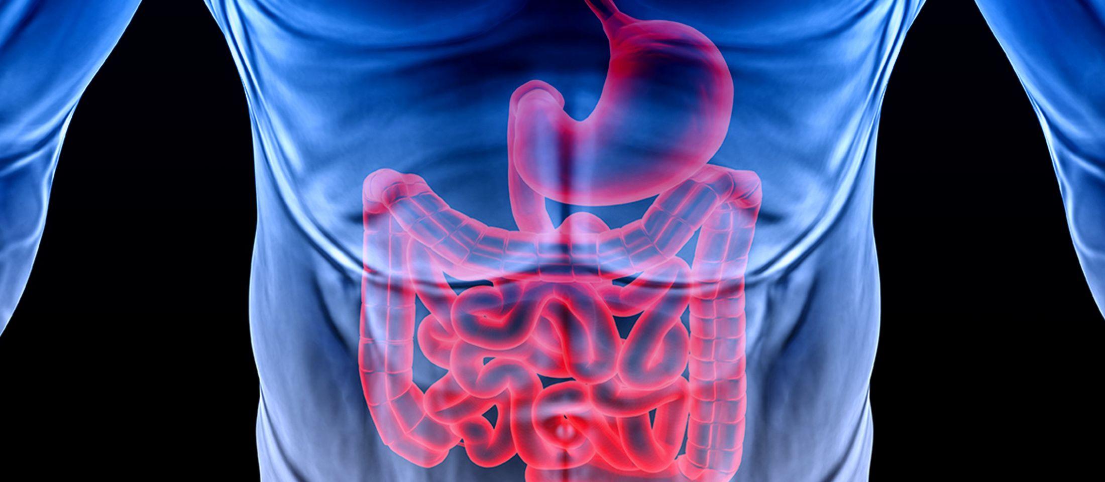
The activities of the Department of Gastroenterology at the Hospital cover the whole range of modern Gastroenterology and Interventional Endoscopy.
The Department is directly supported by the Hospital’s Diagnostic-Imaging and Pathology Labs for the conduction of all necessary endoscopic, lab, imaging and histology tests.
Endoscopy Laboratory
The Lab has top-notch equipment such as a Full High Definition endoscopy tower and high-resolution video-endoscopes using the latest techniques in Chromoendoscopy and Νarrow Βand Ιmaging for higher diagnostic accuracy.
Diagnostic upper GI (gastroscopies and duodenoscopies) and lower GI endoscopies (colonoscopies and capsule endoscopies) are performed.
Upper and lower GI interventional endoscopies
- Dilation of stenosis in the esophagus-stomach-bowels, either with bougie- or balloon-dilation.
- Stent placement in stenoses of the esophagus-duodenum-colon. Emergency stent placement in colonic ileus (preoperatively performed to serve as a bridge with the surgery: reduction of morbidity – shorter hospital stay – enterectomy and end-to-end anastomosis, all performed at the same time).
- Endoscopic treatment of esophageal achalasia
- Foreign body removal from the upper and lower GI.
- Management of varicose- or non varicose-related bleedings present in the upper of lower GI, utilizing the full spectrum of hemostatic techniques (infusions of adrenaline-acrylic adhesives-firming agents / Argon Plasma Coagulation (APC) thermopexy / bipolar or monopolar electrosurgery / clips placement / variceal banding.
- Esophageal varices banding: chronic and preventive treatment
- Mucosal resection of early neoplasms and precancerous lesions (mucosectomy)
- APC-assisted endoscopic therapy of gastric antral vascular ectasia syndrome (GAVE-Watermelon stomach)
- Upper and lower GI polypectomies
ERCP- Endoscopic retrograde cholangiopancreatography
- Diagnostic (though endoscopic ultrasonography (EUS) and magnetic resonance cholangiopancreatography (MRCP) are preferred on safety as well as on precision grounds)
- Interventional ERCP (sphincterectomy- stones removal-lithotripsy- placement of plastic and metallic stents with auto-dilating capacity-placement of drainage catheters)
Endoscopic Ultrasonography (EUS)
What is an Endoscopic Ultrasound?
An Endoscopic Ultrasound (EUS) is method combining endoscopy with ultrasounds.
It is performed with special endoscopic instruments which have an ultrasound head at the one end. During upper or lower GI tract endoscopy the assistance of the ultrasounds enables us to view the outer lumen of the tract. Therefore, we can thoroughly study the wall structure of the GI tract as well as all the details of tissues and organs adjacent to the tract, such as pancreas, liver, spleen, biliary ducts, lymph nodes and vessels. What is the advantage of the method?
The advantage lies within the ability to place the ultrasound head very closely to the organs and tissues we want to study, as it delivers high-resolution images. Thus, the Endoscopic Ultrasound can well-define and characterize any findings from other imaging tests, such as CT-scan or MRI-scan, with high precision, and in a number of cases detect very small lesions, measuring only millimeters, which are not detectable by traditional imaging methods.
Fine needle aspiration and Doppler
With the Endoscopic Ultrasound we can check the vascular perfusion (Doppler) as well as obtain specimen from the lymph nodes, and suspicious masses with the assistance of an ultrasound-guided fine needle. The collected specimen can then be studied by a specialized Cytologist for the establishment of the diagnosis. The procedure is called Fine Needle Aspiration-FNA, and compared to other methods, it is less invasive and safer.
Cutting-edge technology – Elastography
Metropolitan General’ state-of-the-art Endoscopic Ultrasonography equipment has the capacity of conducting Elastography, i.e. a pioneering technique which helps us evaluate the hardening of the examined tissues.
It is known that some conditions, such as cancer, can result in tissue hardening. Elastography, a technique which allows the evaluation of tissue hardening during an Endoscopic Ultrasound, provides us with this additional information which can serve in the diagnosis. With the assistance of Elastography, we can guide the fine biopsy needle to the most hardened part of the lesion we study and thus, maximize the likelihood of a successful test.
How is the Endoscopic Ultrasound performed?
In the majority of cases, the Endoscopic Ultrasound is performed with the patient lightly sedated (i.e. drowsy but still able to be woken). In this way, the patient does not feel any discomfort and the biopsy procedure is painless. Most of the times, the patient can go home on the same day after the procedure while the results are readily available. However, in case a biopsy specimen has been collected, there may be a slight delay until the results of the cytology examination are delivered.
Diagnostic Applications
- Diagnosis and staging of tumors present in the esophagus, pancreas and rectum
- Lung cancer staging
- Chronic pancreatitis assessment
- Pancreatic masses and cysts evaluation
- Study of biliary conditions, such as lithiasis or tumors in the gall bladder, biliary ducts or liver
- Study of the anal sphincters as part of the investigation for urinary incontinence
- GI submucosal tumors investigation
- Enlarged lymph nodes study
Therapeutic applications
- Cysts and abscesses drainage
- Abdominal mesh neurolysis (in cases of surgically untreatable pancreatic cancer, as part of pain adjuvant treatment)
ESD: ENDOSCOPIC SUBMUCOSAL DISSECTION
What is the ESD technique?
Endoscopic Submucosal Dissection (ESD) is an innovative interventional endoscopy technique that originated from Japan with clinical experience of over twenty years.
ESD is the most recent development in endoscopic interventional techniques that allow en-block (total) excision of the largest and histologically advanced epithelial lesions, including early cancer in the upper and lower gastrointestinal tract (esophagus, stomach, large intestine) and a wide range of mucosal lesions, that until recently required surgical removal.
Through the ESD technique the lesion area in the digestive tract is removed with utmost precision, the organ’s muscle layer is left untouched and therefore surgical excisions of ailing organs are avoided.
What digestive lesions are treated with ESD?
The digestive tract wall (esophagus, stomach, large intestine) is constituted by various layers that include (from the inside outwards) the mucosa, the submucosal layer, the muscle layer and the serosa.
All digestive system lesions start to appear in the mucosa and especially the large intestine may present the so-call polyps. In the esophagus and the stomach the initial damages are usually not clearly detectable. Then dysplastic problems start appearing in the mucosal cells that range for low- to high-degree lesions. The dysplastic lesions then extend beyond the mucosa to the lower layers of the wall such as the submucosal layer, while a malignant penetrative process may go as far as the organ’s muscle or migrate through submucosal layer vessels beyond the wall to the local lymph nodes.
ESD may be performed when the early lesion is located in the mucosa or the upper submucosal layers (stage T1a, T1b1). Endoscopic removal of such lesions is now possible and oncologically sufficient in case the malignant lesion does not go beyond the deeper submucosal layers (T1b2).
In order to assess whether a lesion can be endoscopically removed, an endoscopic evaluation is required with high-resolution magnification endoscopy, chromo-endoscopy and/or ultrasound endoscopy. Latest generation video endoscopy systems offer Image Enhanced Endoscopy, Magnification Narrow-Band Imaging (IEE/ME-NBI). These technological breakthroughs guarantee prompt diagnosis of malignant lesion in the esophagus, the stomach and the large intestine at a very early stage during a gastroscopy or colonoscopy procedure.
In reality only a endoscopy review may give us the opportunity of a “visual” biopsy of a lesion at a 85× magnification scale of the actual size so that an endoscopy physician may confirm the kind of problem, receive a targeted biopsy or perform an endoscopic excision within healthy peripheral and vertical boundaries and deeper level lesion dissection, thanks to ESD method.
How were these lesions treated before ESD?
Until recently the usual practice in endoscopic detections of such lesion consisted in endoscopic biopsy. On the basis of the results of such a process a new endoscopic removal was scheduled by means of a diathermy snare to one or more parts (Piecemeal EMR – Endoscopic Mucosal Resection) or partial or total surgical excision of the organ.
The waiting period for the endoscopic biopsy results and a possible formation of fibrosis on the wall (especially in cases of damages in the large intestine) made prospective endoscopic therapies very harsh and ineffective.
Even in cases of endoscopic removal frequently pathological tissue would remain in the organ or the lesion could extend to deeper layers causing relapse rates of 21% to 46% of the total cases. The patient was then led to repeated endoscopic excisions or open surgery.
How effective is ESD?
The ESD technique offers:
- Terminal removal of malignant lesions in 95% in stomach conditions and 92.5% in large intestine cases.
- Almost zero rates of relapse (between 0 and 1% of all cases).
- The oncological philosophy of ESD allows precise histopathological staging and analysis of malignant tissues, since the lesion is removed from tissues at maximum depth margins part while offering the safest future monitoring of patient with R0 excision or establishing succinct indications for oncology surgery in cases of massive profound penetrations into the submucosal layer (T1b2 stage).
ESD vs. EMR (conventional Endoscopic Mucosal Resection)
In comparison with conventional Endoscopic Mucosal Resection (EMR), typically used in large intestine superficial polyps, regardless of lesion size and location, ESD is more effective than successful en-block excisions by a rate of 95% over 58%, by a rate of 89% over 22% for treatment excision (R0) and by a rate of 0.3% vs. 25% for localized relapses.
On the basis of the foregoing rates, ESD has been demonstrated to be more clinically effective than EMR and minimally interventional procedures (compared with surgical resections). Although hemorrhage and perforation may be noted at rates of 9% and 4.5% respectively (for EMR 5.8% and 1% respectively) of all cases, these conditions are treated effectively at the time of operation with no health condition consequences.
The European Society of Gastrointestinal Endoscopy (ESGE) has proposed since 2015 that all early stomach and esophagus lesions be removed with ESD. For large intestine cases ESD is proposed in particular polyps types and almost in all cases of rectal polyps measuring more than 2.5 cm.
How is ESD performed?
ESD is an endoscopic surgical method and is performed with the help of an endoscope instrument inserted through the mouth or the rectum depending on the location of the detected tumor (at the esophagus, the stomach or the large intestine). Throughout the entire course of the procedure the patient is monitored by an anesthesiologist for the maintenance of sedation.
This procedure includes the following stages:
- Thermal coagulation current marking of the healthy limits around the lesion perimeter under direct high-definition endoscope probe review following the marking of the lesion with chromoendoscopy.
- Infusion of special solution into the submucosal layer with a sclerotherapy needle.
- Total peripheral mucosal incision with a special electrosurgical scalpel.
- Dissection of the mucosal-submucosal margins.
- Total dissection along the entire width and the length of the submucosal layer and en-block (total) excision of the neoplasm.
- Review of the artificial ulcer and prophylactic hemostasis of vessels visible in the muscle layer.
- Fixing the lesion on a special cork board and preparations for a histological examination.
The basic precondition for safe ESD application to all patients is the education of the endoscopic specialists at renowned centers known for their solid experience (especially in Asia and Europe) as ESD is a specially challenging technique.
Additionally, the presence of the proper latest-generation endoscope probe equipment is necessary offering the possibility of high-resolution enhanced images and surgical features.
Advanced endoscopic interventional procedures such as ESD are carried out in specialized endoscopic facilities applying absolute safety and effectiveness standards to all patients.
Metropolitan General is one of the few internationally recognized treatment centers applying this particular technique under the direction of Dr Stefanos Basioukas, Gastroenterologist and Interventional Endoscopy Specialist, Director of Advanced Clinical Endoscopy.
Our Clinics
1st Gastroenterology Clinic
2nd Gastroenterology Clinic of Invasive Endoscopy
3rd Gastroenterology - Hepatology Clinic
4st Gastroenterology Clinic
5th Gastroenterology Clinic of Interventional Endoscopy
Gastroenterology Clinic of Invasive Endoscopy
Gastroenterology Clinic of Advanced Therapeutic Endoscopy
Gastroenterology - Hepatology Clinic
Contact Numbers: +30 210 6502624, +30 210 6502118


