


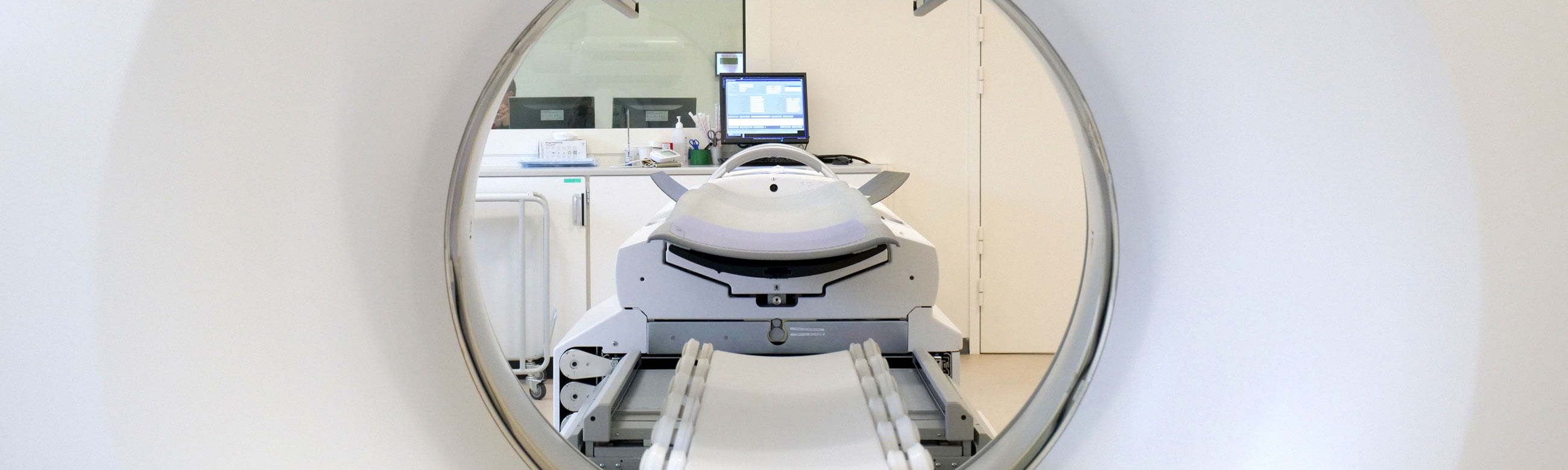




The Nuclear Medicine Laboratory includes an in vivo studies unit where scintigraphy examinations are performed.
Scintigraphy examinations provide both morphological and functional assessment of organs using special substances known as radiopharmaceuticals, which are administered intravenously. The radiopharmaceutical contains a small amount of radioactive isotope combined with medications specific to the organ being examined. These examinations are safe, and the radiation dose is so low that it poses no danger to either the patient or the surrounding environment.
Scintigraphy Scans performed at the Department:
- Myocardial Perfusion Imaging (MPI): Stress and Rest studies, including Gated SPECT analysis
- Thyroid Scintigraphy (Thyroid Scan)
- Parathyroid Scintigraphy (Parathyroid Scan)
- Renal Scintigraphy: Static (DMSA) and Dynamic (DTPA/MAG3) studies
- Bone Scintigraphy: Whole-body and Three-Phase Bone Scan
- Lung Scintigraphy: Perfusion and Ventilation (V/Q Scan)
- Brain Scintigraphy:
- Brain Perfusion SPECT
- DaTscan (Dopamine Transporter Imaging)
- Radionuclide Cisternography (CSF Flow Study)
- Gastrointestinal (GI) Bleeding Scintigraphy
- Gastric Emptying Study
- Lymphoscintigraphy: Sentinel Lymph Node (SLN) Mapping/Localization
- Equilibrium Radionuclide Angiocardiography (ERNA): Multigated Acquisition (MUGA) Scan at rest
The Nuclear Medicine Department features a fully equipped Hot Lab and a shielded waiting area for patients undergoing examination.
Operating Hours: Monday-Friday, 08:00-16:00
Contact number: +30 210 650 2569
Gamma Camera
After administration, the radiopharmaceutical accumulates in the target area, emitting radiation that is then captured by a specialized imaging device known as a Gamma Camera (SPECT).
Unlike conventional Radiology, where the machine emits radiation that passes through the patient, Nuclear Medicine utilizes "open sources" of radiation. In this case, it is the patient who temporarily emits information, which is then collected and processed by the Gamma Camera.
Metropolitan General is equipped with the state-of-the-art Siemens Symbia Evo Excel Gamma Camera system. This advanced unit is designed for static, dynamic, whole-body, and tomographic nuclear medicine studies.
- Enhanced Diagnostics: Features superior image quality for improved detection and precise lesion characterization, reducing the need for additional testing.
- Patient-Centric Design: Its ergonomic structure enhances the examination experience for bariatric patients, individuals with mobility issues, or those suffering from claustrophobia.
Myocardial Perfusion Imaging
Myocardial Perfusion Imaging (MPI) is a Nuclear Medicine study that evaluates the blood flow to the heart muscle during both stress and rest. It involves the intravenous administration of a radioisotope (radiopharmaceutical) that "illuminates" the myocardium for imaging.
Clinical Utility
- Identifies Ischemia (blood flow issues during stress)
- Detects Myocardial Scarring from previous infarctions (heart attacks)
- Assesses Myocardial Viability
Procedure
- Injection of a small amount of radiopharmaceutical
- Rest Phase: Imaging with the Gamma Camera
- Stress Phase: Exercise on a treadmill or pharmacological stress
- Second injection and Stress Phase imaging
Interpretation of results
- Normal: Uniform blood flow/perfusion
- Ischemia: Perfusion defect observed only during the stress phase
- Infarct (Scar): Perfusion defect present in both stress and rest phases
Duration: Approximately 2-4 hours in total.