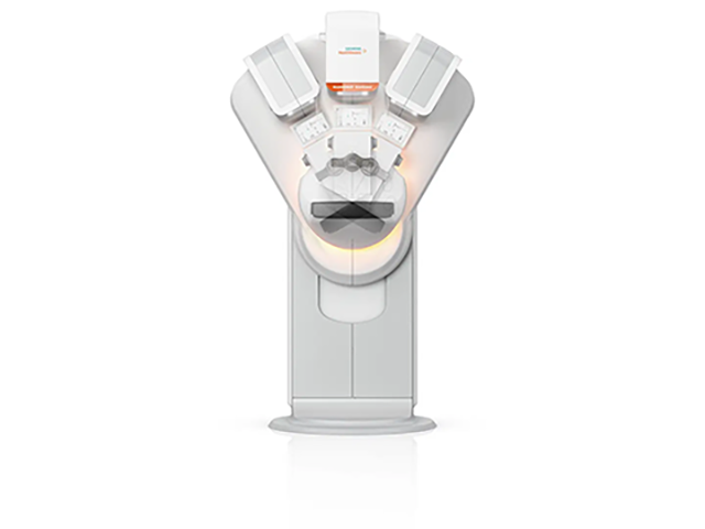
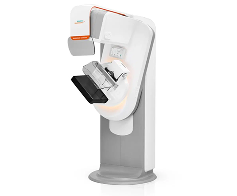
Mammomat revelation mammographic system

The Breast Center at Metropolitan General is the only treatment unit in Greece featuring the worldwide leading Mammomat Revelation mammographic system made by Siemens Healthineers, which performs digital mammography, tomosynthesis, reconstitutive 2D and 3D images, contrast-medium mammography, stereotactic biopsy and tomosynthesis biopsy. It also features pioneering technical features that profoundly improve the entire diagnostic outcome through a reduced radiation dose up to 30%.

  • Wide-angle 3D breast tomosynthesis. Mammomat Revelation is the sole system with a 50° scanning angle, the widest scanning angle in the world (in contrast to other tomosynthesis devices with much more limited scanning angles, usually 15°). This presents an important difference since wide-angle scans provide better and more detailed geometric information that may be used in 3D reconstitution. In total, we may use 25 images in this particular system in order to attain 3D imaging, thus resulting in the greatest possible resolution ability and the best possible identification of the various breast structures. This leads to maximum diagnostic safety, increasing the possibility of localizing even the least traceable breast lesions, and reducing the false positive/negative results and follow-ups for supplementary controls.
  • The innovative Prime technology allows, under certain preconditions, reduction in radiation doses by up to 30% combined with the possible images.
  • Ability of reconstitutive Insight 2D scans which feature images comparable to conventional digital mammography. Thus, we can compare the tomosynthesis control with other prior conventional digital mammographic scans, without the use of any additional conventional scans (thus reducing radiation doses by up to 40%).
  • Insight 3D imaging that allows the creation of new actual easy-to-understand 3D video recordings.
  • Automated breast density assessment allowing immediate planning or modification of breast imaging strategy in the course of the examination session on the basis of breast density. Any increased density is regarded as a significant risk factor for breast cancer, while in all recent international medical directives all patients must also undergo a tomosynthesis scan.
  • Automated personalized breast compression: This reduces the intensity and the duration of compression resulting in minimization of patient pain and discomfort. Breast compression is necessary for a sound diagnostic outcome and radiation dose reduction. The greater the compression, the better the tissue identification, the better the mammogram resolution. The thinner the breast thickness, the lesser the radiation dose.
  • TiCEM contrast-medium mammography. This kind of mammography may be used as a direct diagnostic tool in relation with MRI scanning in women with diagnostic problems, family medical history and inability to perform an MRI mammogram.
  • Pioneering stereotactic localization and breast biopsy with/without tomosynthesis (HD tomobiopsy). This allows us to focus with +/- 1 mm precision, using wide-angle tomosynthesis (wide-angle technology). Thus, we attain the best precision and the greatest reduction in procedure time. The procedure is conducted in a comfortable specialized biopsy chair under local anesthesia and a miniscule incision of just a few mm. The innovative InSpect Imaging Tool gives us the opportunity for real-time biopsy tissue radioscopy on the same device, thus improving the interventional procedure technique and, for the same reason, the overall comfort of the examination.

Μηχάνημα Εξέτασης Μαστού