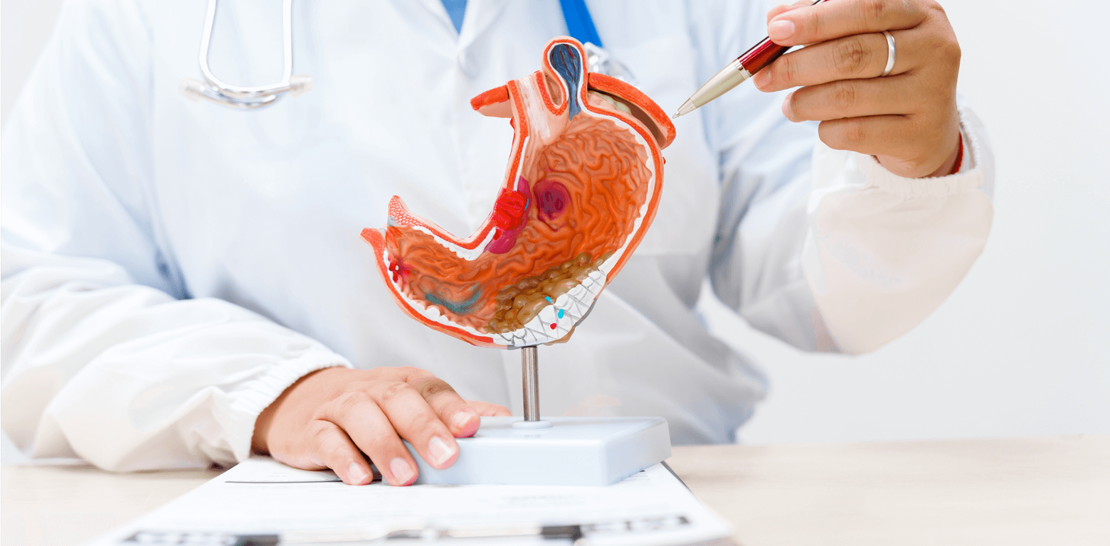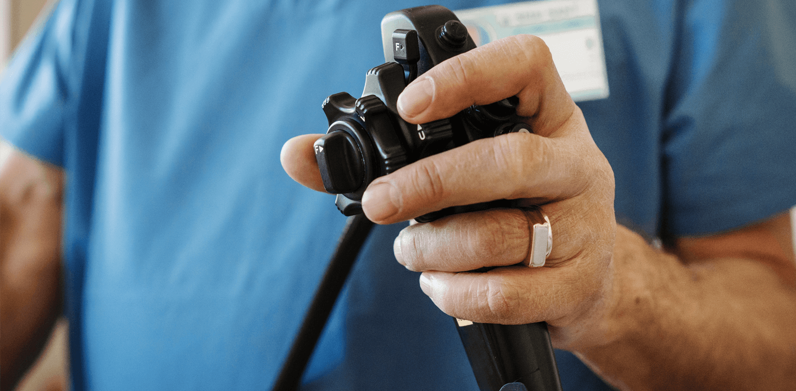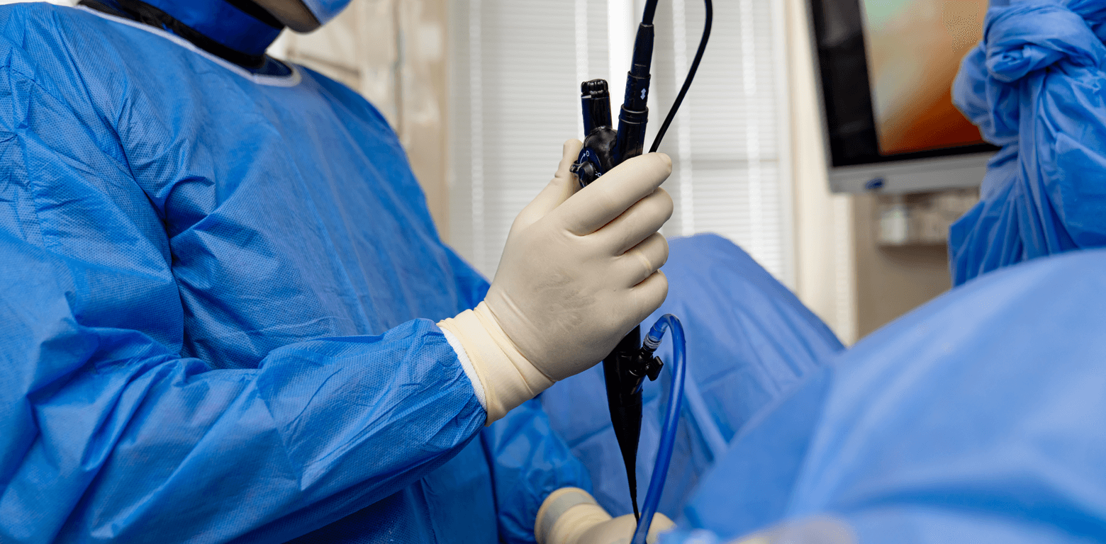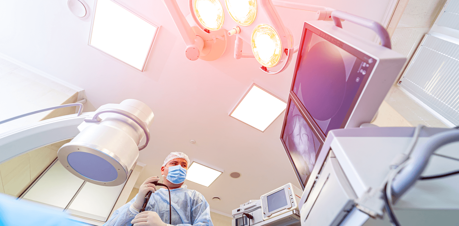




The activities of the Gastroenterology Department of Metropolitan General cover the entire spectrum of modern Gastroenterology and Interventional Endoscopy.
The Gastroenterology Department is directly supported by the Clinic’s Diagnostic-Imaging Laboratories and the Pathology Department for the performance of all required endoscopic, laboratory, imaging, and histological examinations.
Endoscopy Laboratory
The Endoscopy Laboratory is equipped with state-of-the-art technology, including a full high-definition endoscopic tower and high-resolution video endoscopes applying the latest Chromoendoscopy and Narrow Band Imaging techniques for higher diagnostic accuracy.
Diagnostic endoscopies of the upper gastrointestinal tract (gastroscopies and duodenoscopies) and of the lower gastrointestinal tract (colonoscopies and capsule endoscopy) are performed.
Interventional Endoscopy of the Upper and Lower Gastrointestinal Tract
- Dilatation of strictures of the oesophagus, stomach and intestine using either bougies or balloon dilators
- Placement of stents in strictures of the oesophagus, duodenum and large bowel. In addition, emergency stent placement in large bowel ileus (preoperatively as a bridge to surgery: reduction of morbidity - reduction of hospital stay - enterectomy and end-to-end anastomosis in a single stage)
- Endoscopic treatment of oesophageal achalasia
- Removal of foreign bodies from the upper and lower gastrointestinal tract
- Management of upper and lower gastrointestinal bleeding of both variceal and non-variceal aetiology with a full range of haemostatic techniques (adrenaline injection-cyanoacrylate glue-sclerosants / Argon Plasma Coagulation (APC) / bipolar or monopolar electrocoagulation / clip placement / variceal band ligation)
- Oesophageal variceal ligation (banding): chronic and prophylactic treatment.
- Endoscopic mucosal resection of early neoplasms and precancerous conditions (mucosectomy)
- Endoscopic treatment of radiation colitis - gastric antral vascular ectasia (GAVE – Watermelon stomach) with APC
- Polypectomies of the upper and lower gastrointestinal tract
ERCP - Endoscopic Retrograde Cholangiopancreatography
ERCP is performed by specialised interventional endoscopists in the presence of an anaesthesiologist in a dedicated fluoroscopy suite of the Clinic.
Metropolitan General is equipped with a latest-generation, fully digital, remote-controlled fluoroscopy system with a flat-panel detector, providing optimal image quality and high spatial resolution with the lowest possible radiation dose.
- Diagnostic (although Endoscopic Ultrasound (EUS) and Magnetic Resonance Cholangiopancreatography (MRCP) are preferred due to higher safety and accuracy)
- Interventional ERCP (sphincterotomy - stone extraction - lithotripsy - placement of plastic and self-expandable metal stents – placement of drainage catheters)
EUS - Endoscopic Ultrasound
What is Endoscopic Ultrasound?
Endoscopic Ultrasound (endoscopic ultrasound or EUS) is a method that combines endoscopy with ultrasound.
It is performed using specialized endoscopes that have an ultrasound transducer at their tip. During endoscopy of the upper or lower gastrointestinal tract, with the aid of ultrasound we are able to visualize beyond the lumen of the gastrointestinal tract. Therefore, we can study in great detail both the structure of the wall of the gastrointestinal tract and the tissues and organs adjacent to it, such as the pancreas, liver, spleen, biliary system, lymph nodes and blood vessels.
Where does the method excel?
The ability to position the ultrasound probe very close to the organs and tissues under examination provides images of exceptionally high resolution. Thus, Endoscopic Ultrasound can accurately clarify and characterize findings from other imaging modalities, such as computed tomography or magnetic resonance imaging, and can often detect small lesions of millimeter size that are not visible with conventional imaging methods.
Needle biopsy and Doppler
With Endoscopic Ultrasound, vascular flow can be assessed (doppler), and samples can be obtained from lymph nodes and suspicious masses using a fine needle under ultrasound guidance. The collected material can subsequently be analyzed by a specialized cytopathologist for diagnostic purposes.
This procedure is termed Fine Needle Aspiration (FNA) and, compared with other methods, is less invasive and safer.
State-of-the-art technology - Elastography
The modern Endoscopic Ultrasound system at Metropolitan General includes elastography, an advanced technique that allows assessment of tissue stiffness.
It is known that certain conditions, such as cancer, lead to increased tissue stiffness. Elastography enables evaluation of tissue stiffness during Endoscopic Ultrasound, providing additional information that may assist in diagnosis. With elastography, the biopsy needle can be guided to the stiffest part of the lesion under examination, thereby maximizing the likelihood of diagnostic success.
How is Endoscopic Ultrasound performed?
Endoscopic Ultrasound is performed in the majority of cases under sedation. Thus, the examination is comfortable for the patient and the biopsy procedure is painless.
In most cases, the patient can return home on the same day after the end of the examination, and the results may be available immediately. However, in cases where a biopsy has been obtained, there is a short delay of a few days until completion of the cytological examination.
Diagnostic Applications
- Diagnosis and staging of cancers of the oesophagus, stomach, pancreas and rectum
- Staging of lung cancer
- Assessment of chronic pancreatitis
- Assessment of pancreatic masses and cysts
- Study of diseases of the biliary system, such as lithiasis or tumors of the gallbladder, bile ducts or liver
- Study of the anal sphincters in the investigation of incontinence
- Study of submucosal tumors of the gastrointestinal tract
- Study of enlarged lymph nodes
Therapeutic Applications
- Drainage of cysts and abscesses
- Celiac plexus neurolysis (in cases of inoperable pancreatic cancer as palliative treatment of pain)
ESD - Endoscopic Submucosal Dissection
What is the ESD technique?
Endoscopic submucosal dissection (ESD) is an innovative interventional endoscopic technique originating from Japan, with more than 20 years of clinical experience.
ESD represents the latest advancement in interventional endoscopic techniques, allowing en-block resection (in a single piece) of larger and histologically advanced epithelial lesions, including early cancer of the upper and lower gastrointestinal tract (esophagus, stomach, colon), as well as a wide range of mucosal lesions that previously required surgical removal.
With the ESD technique, the lesion area in the gastrointestinal tract is removed with high precision, while the muscular layer of the organ remains intact, thus avoiding surgical resection of the affected organ.
In which gastrointestinal lesions is ESD applied?
The wall of the gastrointestinal tract (esophagus, stomach, colon) consists of multiple layers, including (from inner to outer) the mucosa, submucosa, muscularis propria, and serosa.
All gastrointestinal lesions initially appear in the mucosa and, particularly in the colon, form what is known as a polyp. In the esophagus and stomach, early lesions are usually not clearly distinguishable. Subsequently, dysplastic changes develop in mucosal cells, ranging from low- to high-grade. These dysplastic lesions then extend beyond the mucosa into deeper layers of the wall, such as the submucosa, where malignant invasive transformation begins, potentially progressing into the muscular layer of the organ or, via submucosal vessels, beyond the wall into regional lymph nodes.
ESD can be performed when the early lesion is confined to the mucosa or the superficial layers of the submucosa (stage T1a, T1b1). Endoscopic removal of such lesions is feasible and oncologically adequate when the malignant lesion does not extend beyond the deeper layers of the submucosa (T1b2).
To determine whether a lesion can be removed endoscopically, it must be evaluated using high-definition magnifying endoscopy, chromoendoscopy, and/or endoscopic ultrasound. Latest-generation video endoscopic systems incorporate image-enhanced endoscopy technology (IEE/ME-NBI - Image Enhanced Endoscopy, Magnification Narrow Band Imaging), ensuring early detection of malignant lesions in the esophagus, stomach, and colon at a very early stage during gastroscopy or colonoscopy.
Essentially, optical biopsy under endoscopic visualization at up to 85× magnification allows the endoscopist to assess the nature of the lesion and obtain targeted biopsies or proceed with endoscopic resection with clear lateral and vertical margins through deeper-layer dissection using the ESD method.
What was previously done for these lesions?
Until recently, when a lesion was detected during endoscopy, the standard approach involved endoscopic biopsy, followed by resection planning based on the result, either via endoscopic removal using a diathermic snare in one or more pieces (piecemeal EMR) or surgical resection of part or the entire organ.
Waiting for biopsy results and the possible development of fibrosis in the wall, particularly in colonic lesions, often made subsequent endoscopic treatment difficult and ineffective.
Even in cases of endoscopic removal, residual pathological tissue often remained, or the lesion extended into deeper layers, leading to recurrence rates of 21–46%. Patients were then subjected to repeated endoscopic resections or open surgery.
How effective is ESD?
The ESD technique offers:
- Definitive removal of malignant lesions in 95% of cases in the stomach and 92.5% in the colon.
- Near-zero recurrence rates (0–1% of cases).
- The oncological principle of ESD allows precise histopathological staging and analysis of malignant tissue, as the lesion is removed in a single piece with maximal deep margins. It provides the safest approach for future surveillance in cases of R0 resection or enables clear indications for oncologic surgery in cases of deep submucosal invasion (stage T1b2).
ESD vs EMR (conventional endoscopic mucosal resection)
Compared with conventional endoscopic mucosal resection (EMR), which is mainly used for superficial colonic polyps, ESD, regardless of lesion size and location—demonstrates superiority in en-block resection (95% vs 58%), curative (R0) resection (89% vs 22%), and local recurrence (0.3% vs 25%), respectively.
Based on these outcomes, ESD is clinically more effective than EMR and minimally invasive compared to surgical treatment. Although it is associated with bleeding and perforation rates of 9% and 4.5% (compared to 5.8% and 1% for EMR), these are effectively managed during the procedure without clinical consequences.
Since 2015, the European Society of Gastrointestinal Endoscopy (ESGE) recommends that all early lesions of the stomach and esophagus be removed using ESD. For the colon, ESD is recommended for specific types of polyps and for most rectal polyps larger than 2.5 cm.
How is ESD performed?
ESD is an endoscopic-surgical method performed using an endoscope inserted through the mouth or rectum, depending on the tumor location (esophagus, stomach or colon). The patient is continuously monitored by an anesthesiologist throughout the procedure for sedation.
The procedure includes the following stages:
- Marking the lesion margins with coagulation current on healthy tissue under direct visualization using a high-definition endoscope after delineation with chromoendoscopy.
- Injection of a special solution into the submucosal layer using a sclerotherapy needle.
- Circumferential mucosal incision using a specialized electrosurgical knife.
- Dissection along the mucosa-submucosa interface.
- Complete dissection across the full width and length of the submucosal layer and en-block resection of the neoplasm.
- Inspection of the artificial ulcer and prophylactic hemostasis of visible vessels on the muscular layer.
- Fixation of the specimen on a special cork board and preparation for histological examination.
Safe application of ESD requires that the endoscopist be trained in high-expertise centers (mainly in Asia and selected European centers), as it is a highly demanding technique.
It also requires the availability of advanced, state-of-the-art endoscopic equipment with high-resolution and image-enhancement capabilities, as well as interventional features.
Advanced endoscopic procedures such as ESD are performed in specialized endoscopy units with a focus on maximum safety and effectiveness for the patient.
Metropolitan General is among the few centers worldwide where this technique is performed by Mr. Stefanos Basioukas, Gastroenterologist - Interventional Endoscopist, Director of the Advanced Therapeutic Endoscopy Clinic.