


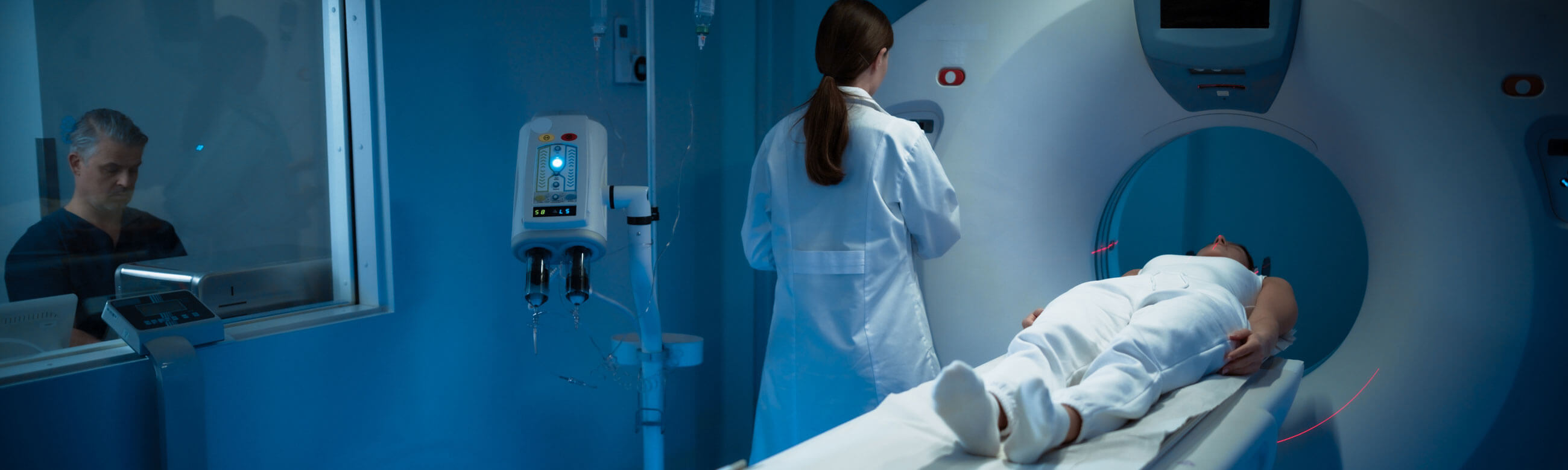




The Imaging Laboratories are one of the most important pillars of our Clinic, providing comprehensive, high-precision diagnostic services. Their aim is the timely and reliable imaging of the body’s organs and tissues, contributing decisively to accurate diagnosis and the effective therapeutic management of each case.
With advanced technological equipment and specialized personnel, the laboratories cover the full spectrum of imaging examinations, including digital radiography, CT and MRI scans, digital mammography, bone mineral density testing, and specialized angiographic procedures. All examinations are performed in a safe and comfortable environment.
Director: Kaimenakis Dimitrios, Radiologist
Contact numbers: +30 210 650 2510-2511
Diagnostic Radiology Department
The Diagnostic Radiology Unit of Metropolitan General is a premier, fully equipped facility in Greece, dedicated to providing high-quality medical imaging services. We combine safety, reliability, and rapid results to support every patient’s healthcare journey. Equipped with cutting-edge medical technology and staffed by highly experienced radiologists and technicians, our department covers the full spectrum of diagnostic examinations. We specialize in high-precision imaging that is fundamental to early detection and accurate clinical diagnosis.
All diagnostic tests are performed in a comfortable and friendly environment. Our priority is to ensure a stress-free experience for every patient while maintaining the highest standards of imaging quality.
Through continuous medical training and strict adherence to international quality protocols, we ensure the highest reliability in diagnostic reporting. Our department works in seamless collaboration with all hospital units, offering an integrated diagnostic approach for targeted and effective treatment plans.
Digital Radiology
The Radiology Laboratory of Metropolitan General is fully equipped with state-of-the-art digital technology, ensuring superior imaging quality, significantly reduced radiation dose, and the elimination of repeat examinations.
A full range of radiological procedures is performed, from standard digital X-rays to specialized radiographic and fluoroscopic studies, such as intravenous pyelography, barium meal studies, enteroclysis, and fistulography.
The Department is equipped with a latest-generation, fully digital, remote-controlled fluoroscopy system featuring a flat-panel detector, providing optimal image quality and high spatial resolution with the lowest possible radiation exposure.
In addition, it includes a digital radiography system with an advanced ortho package, enabling full-length imaging of the spine and lower extremities.
Modern Digital Archiving and Processing of Medical Images
The Radiology Laboratory has a fully developed Picture Archiving and Communication System (PACS) for the management and storage of medical images. Once the examination is completed, the images are stored and managed electronically, separately for each patient.
The use of high-resolution monitors, post-processing capabilities, and access to previous examinations for comparison allow the imaging physician to obtain a comprehensive view of the patient’s findings and provide faster, safer diagnosis.
Operating Hours: Monday–Friday, 07:00–23:00 & Saturday, 08:00–16:00
Emergency coverage: 24/7
Contact numbers: +30 210 650 2510-2511
Orthopantomography
Excellent Imaging Quality with the Lowest Possible Radiation Dose
Metropolitan General is equipped with the latest-generation Orthophos XG 3D Ready digital orthopantomograph by Sirona, ensuring highly reliable, safe, and rapid panoramic and cephalometric imaging of the teeth and jaws.
With a wide range of advanced diagnostic programs, this panoramic imaging system covers the dental needs of every patient, including endodontics, implantology, periodontology, orthodontics, and oral surgery.
Its main advantages include:
- Excellent image quality and enhanced diagnostic accuracy, even in complex cases, through the innovative CsI sensor and ASTRA algorithm
- Very low radiation dose
- Faster image acquisition and avoidance of incorrect imaging and unnecessary radiation exposure, through the EasyPad control panel combined with automatic patient positioning
- Safe and immediate patient positioning through the specialized Bite Block, which determines the optimal head inclination and helps maintain a stable position
Operating Hours: Monday–Friday, 07:00–23:00 & Saturday, 08:00–16:00
Contact numbers: +30 210 650 2510-2511
Bone Mineral Density Measurement
Bone Mineral Density measurement (BMD), also known as bone densitometry, is an imaging examination that assesses how dense and strong the bones are. It is mainly used for the diagnosis of osteoporosis and assessment of future fracture risk.
The examination is performed using Dual-Energy X-ray Absorptiometry (DEXA). The patient lies on a specialized scanner, while a very low dose of X-rays measures the amount of calcium and other minerals in the bones. The results help the physician assess fracture risk and recommend the appropriate therapeutic approach.
At Metropolitan General, we use a modern, innovative Fan-Beam technology system. This technology increases diagnostic information and accuracy while ensuring the lowest possible radiation dose, reducing exposure by up to tenfold. Bone mineral density measurement is performed with the DEXA method in the following areas:
- Lumbar spine
- Hip
- Forearm
- Whole body
Bone mineral density testing is indicated for:
- Perimenopausal women
- Women after total hysterectomy
- Amenorrhea or oligomenorrhea
- Prolonged immobilization
- Long-term corticosteroid therapy
- Hyperthyroidism or long-term thyroxine treatment
- Evaluation of treatment outcomes
- Men with hypogonadism or radiological evidence of osteopenia
- Medical conditions such as renal failure, liver failure, and hemolytic anemias
- Malignancies, including multiple myeloma and Paget’s disease
- Hereditary risk factors
Read more about: Osteoporosis Unit
Operating Hours: Monday-Friday, 07:00-15:00
Contact number: +30 210 650 2569
Digital Mammography with Tomosynthesis
Mammomat B.brilliant: A Leading Low-Dose Mammography System
The Mammomat B.brilliant mammography system by Siemens Healthineers, available at our Breast Center, is an advanced tool for breast cancer diagnosis, combining speed, high resolution, and innovative technologies.
Mammomat B.brilliant features PlatinumTomo, an innovative wide-angle tomosynthesis technology with a 50° angle, offering the world’s fastest wide-angle tomosynthesis and a scan time of approximately 5 seconds. It produces high-quality 3D images in minimal time.
With the Premia image reconstruction algorithm, which integrates artificial intelligence (AI) applications, image noise is reduced and image quality is optimized while maintaining very low patient dose levels.
Mammomat B.brilliant also offers automatic breast density assessment and features Prime technology, which ensures a low radiation dose.
One of its additional advantages is the ability to perform contrast-enhanced mammography- TiCEM (Titanium Contrast-Enhanced Mammography), using an optimized titanium filter for improved visualization. This supports the detection and assessment of suspicious lesions, helping avoid the additional time and cost of an MRI scan.
The system also features a dedicated personalized breast compression function, adapted to each woman’s anatomy, facilitating positioning during the examination, and providing an improved, more comfortable experience.
Computed Tomography (CT)
The CT Laboratory operates two multislice CT scanners, enabling the performance of all modern CT examinations, including:
- Standard CT scans
- CT angiography of the brain, carotid arteries, aorta, and upper and lower limbs
- Coronary CT angiography
- CT colonography / virtual colonoscopy
- CT enterography - enteroclysis
- Dental CT
- CT pyelography
- Specialized CT examinations using dedicated protocols, such as Signature and other protocols suitable for preoperative orthopedic measurements
During CT examinations, low-dose protocols are applied, including for chest CT scans.
In addition to diagnostic examinations, the CT Laboratory also performs CT-guided interventional procedures, including aspirations, biopsies, drainage of collections, ablation procedures, fiducial placement, and more.
Operating Hours: Monday–Friday, 07:00–21:00 & Saturday, 08:00–14:00
Emergency coverage: 24/7
Contact numbers: +30 210 650 2510-2511
Specialized CT Examinations
- Coronary CT angiography represents a major advancement in primary care. It enables painless and non-invasive evaluation of the coronary arteries and has been shown to be diagnostically equivalent to conventional catheter-based coronary angiography in detecting hemodynamically significant stenoses requiring treatment. The examination is recommended for patients with: a) atypical precordial pain without clear pathological findings in other cardiology tests b) low to moderate probability of coronary artery disease based on laboratory testing and personal/family history c) history of bypass surgery, to assess graft patency d) in selected cases, history of stent placement, to assess stent patency.
- Calcium scoring measures the amount of calcium deposited in the walls of the coronary arteries. This amount has been associated with the likelihood of developing heart disease. The examination can be performed either as a standalone test or as part of coronary CT angiography.
- CT colonography, also known as virtual colonoscopy, enables the diagnosis of colorectal cancer in a painless, non-invasive way with high sensitivity. Its results are comparable to those of optical colonoscopy. It has been included by the American Cancer Society among the recommended screening examinations for early diagnosis of colorectal cancer. It is recommended for individuals over the age of 50 every five years, as an alternative to optical colonoscopy or barium enema. At the same time, CT colonography allows assessment of all organs of the upper and lower abdomen, as in an abdominal CT scan.
- CT pyelography has largely replaced conventional pyelography, as it provides more information for all diagnostic questions concerning the urinary system, such as renal colic, hematuria, and characterization of renal masses.
- CT enteroclysis and CT enterography are used for the diagnosis of small bowel diseases, such as ileus and inflammatory conditions including Crohn’s disease and celiac disease.
- CT angiography of any vessel in the body is performed painlessly and non-invasively within seconds and is considered equivalent to conventional catheter-based angiography for the diagnosis of critical stenoses.
128-Slice Somatom go.Top CT System
Metropolitan General is equipped with the advanced 128-slice Somatom go.Top CT system by Siemens Healthineers, incorporating the latest low-dose technologies, including Tin Filter, Stellar Detector, and SAFIRE.
The system enables high-diagnostic-accuracy cardiac examinations, such as CT angiography, coronary CT angiography, cardiac function analysis, and cardiac vessel imaging.
The Somatom go.Top CT scanner is accompanied by a tablet for examination planning through a mobile workflow, an integrated camera for patient position monitoring, an intercom system, a dual-head injector, and an independent syngo.via image processing and diagnostic workstation. This supports processing of routine examinations, cardiac cases, pulmonary nodule imaging and analysis, virtual colonoscopy, virtual bronchoscopy, and more.
Magnetic Resonance Imaging (MRI)
Magnetic Resonance Imaging (MRI) is a modern, non-invasive imaging examination that uses a strong magnetic field and radio waves to create highly detailed images of the body’s organs and tissues. It does not use radiation, making it particularly safe.
MRI provides high resolution and accuracy, allowing physicians to detect conditions that may not be visible with other examinations, such as CT scans or ultrasound.
At the MRI and Advanced Imaging Laboratory of Metropolitan General, all types of MRI examinations are performed, including:
- Standard MRI scans
- MR angiography
- Breast MRI
- MR enterography
- MR pyelography
- Rectal MRI
- Perineal MRI
- Magnetic Resonance Cholangiopancreatography (MRCP)
- MR arthrography
- Preoperative liver MRI
- Liver iron quantification
- Multiparametric prostate MRI
- Cardiac MRI
The laboratory is equipped with two modern MRI scanners, 1.5 Tesla and 3 Tesla respectively.
Operating Hours: Monday–Friday, 07:00–23:00 & Saturday, 08:00–16:00
Emergency coverage: 24/7
Contact numbers: +30 210 650 2510-2511
Cardiac MRI
The Cardiac MRI Department of Metropolitan General is certified by the European Association of Cardiovascular Imaging (EACVI), making it one of the few departments in Greece and Europe to hold this distinction.
This EACVI Laboratory Accreditation confirms the high level of services, the excellence of infrastructure, the scientific competence of the staff, and the operating protocols followed, in accordance with international standards.
What Is Cardiac MRI?
Cardiac MRI is one of the most important imaging techniques for the diagnosis of cardiovascular diseases. It uses a strong magnetic field and radio waves to create images of the heart and chest. The acquired images are processed using specialized computer software.
In most cases, a contrast agent, gadolinium, is administered intravenously during the examination to provide additional diagnostic information.
Cardiac MRI is a specialized imaging examination of the cardiovascular system, providing high-quality, high-resolution images that enable more detailed assessment of cardiovascular diseases.
It does not expose the patient to radiation, and the contrast agent used has fewer side effects compared with contrast agents used in other imaging methods.
Advantages
- Modern cardiac imaging technique
- Reliable
- Safe / radiation-free
- Painless / non-invasive
- No special preparation required
- Applicable to all patients with cardiovascular diseases*
*Except for patients with non-MRI-compatible pacemakers or defibrillators, contrast agent allergies, or end-stage renal failure.
Cardiac MRI – Indications
- Coronary artery disease: assessment of ischemic lesions, myocardial viability, and hypokinesia; evaluation of myocardial ischemia after pharmacological stress; coronary artery anatomy
- Myocarditis: viral myocarditis - examination of choice; myocardial involvement in rheumatic diseases
- Assessment of myocardial mass and cardiac anatomy through high-resolution imaging
- Anatomy of the great vessels and aortic aneurysms
- Assessment of native and prosthetic valves, with accurate regurgitation quantification and reliable valve planimetry
- Diagnosis of inflammation of the myocardium and great vessels
- Detection of myocardial iron deposition
- Evaluation of cardiac tumors, both primary and secondary
- Monitoring of pericardial diseases
- Quantification of cardiac chamber volumes and contractility (Gold Standard)
- Investigation of causes of heart failure
- Diagnosis and follow-up of cardiomyopathies, including dilated, hypertrophic, non-compaction, Takotsubo, and others
- Evaluation of surgically repaired congenital heart disease in adults
- Assessment of the right heart chambers
Lead Physician: Vidalakis Eleftherios, Cardiologist specialized in cardiovascular imaging
Assessment of Myocardial Ischemia with Cardiac MRI - Stress Perfusion CMR
Assessment of myocardial ischemia with cardiac MRI is based on a rapid protocol that provides information on both myocardial ischemia and myocardial viability. This examination uses a dedicated program available on the MRI scanner at Metropolitan General.
During the examination, myocardial perfusion under stress conditions can be assessed in real time. The principle of this technique is similar to myocardial perfusion scintigraphy; however, because it is performed using magnetic resonance imaging, it does not expose the patient to radiation.
It is more reliable than conventional scintigraphy, as it provides higher-resolution images, and the examination lasts only 25 minutes. In addition, as a cardiac MRI protocol, it not only detects the presence or absence of myocardial ischemia but can also help identify other possible causes of the patient’s symptoms, even when they are not related to myocardial ischemia.
Specialized Assessment of Myocardial Ischemia and Viability
- Reliability: higher diagnostic reliability than scintigraphy, stress echo, and exercise stress testing; equivalent to FFR-coronary angiography
- Speed: 25-minute duration
- Comfort: pharmacological stress
- Safety: no patient exposure to radiation
- Differential diagnosis: distinguishes ischemia from microvascular angina, pericarditis, extracardiac causes of precordial pain, and other conditions
Lead Physician: Vidalakis Eleftherios, Cardiologist specialized in cardiovascular imaging
Myocardial Mapping with Cardiac MRI
A New, Sensitive Method for Detecting Myocardial Injury Following COVID-19
Myocardial mapping with cardiac MRI is an advanced multiparametric mapping technique based on T1, T2, and T2* magnetic relaxation times. It provides high spatial-resolution images, allowing quantification of changes in myocardial tissue composition at every point. These new techniques are highly effective in detecting diffuse cardiac injury, while also helping monitor the progression of myocardial disease by comparing absolute values in sequential measurements. Myocardial mapping therefore enhances the diagnostic accuracy of cardiac MRI, bringing its reliability close to that of conventional myocardial biopsy.
When Is Myocardial Mapping with Cardiac MRI Indicated?
- Myocarditis: diagnosis, follow-up, and confirmation of complete recovery. In 2018, the technique was officially incorporated into the diagnostic criteria for myocarditis
- Myocardial involvement in autoimmune, rheumatic, and systemic inflammatory diseases, such as lupus, rheumatoid arthritis, sarcoidosis, and others
- Characterization of cardiac tumors
- Myocardial involvement caused by HIV
- Amyloidosis and Fabry disease
- Differential diagnosis of myocardial hypertrophic conditions
- Prognosis of coronary artery disease
- Cardiac involvement following COVID-19
Myocarditis and COVID-19
According to international scientific observations, COVID-19 can also affect the heart, causing either myocardial infarction or, more commonly, inflammation of the myocardium, known as myocarditis.
Most importantly, myocardial inflammation is often asymptomatic and subclinical, meaning that the patient may not be aware that the infection has also affected the heart.
With a single examination, it is therefore possible to assess whether coronavirus infection has affected the heart, even in patients who have not experienced cardiac symptoms. This enables timely intervention and helps protect the myocardium from permanent damage that could cause symptoms in the future.
Patients who have recovered from COVID-19 can now undergo cardiac evaluation at Metropolitan General through a dedicated cardiac MRI protocol based on myocardial mapping techniques. This protocol has been specially adapted for former COVID-19 patients by Eleftherios Vidalakis, Cardiologist specialized in cardiovascular imaging.
Contact numbers: +30 210 650 2510-2511
Lead Physician:
Eleftherios Vidalakis, Cardiologist specialized in cardiovascular imaging
Associate:
Savvas Loizos, Cardiologist specialized in cardiovascular imaging
Advanced 3 Tesla MRI Scanner
Highly Detailed and Rapid Imaging
With a strong focus on innovation and cutting-edge medical technology, Metropolitan General is equipped with the high-power, latest-generation 3 Tesla Skyra Magnetom MRI scanner by Siemens, featuring the most advanced programs that enable even the most specialized and innovative examinations.
It is equipped with state-of-the-art software and advanced multichannel coil technology, ensuring exceptional image quality, increased diagnostic accuracy, and maximum examination speed, with scan times reduced by up to 40%.
Specifically, it features the highest number of channels currently available in Greece for imaging of the head, neck, and abdomen (128 channels in total): 64 channels for head and neck imaging, 60 channels for abdominal imaging, and 18 channels for breast imaging. These can be connected simultaneously for whole-body imaging.
As a result, even whole-body MRI scans can now be performed as a routine procedure, while different body regions can be imaged within minutes in a single rapid acquisition, without the need to reposition the patient or change coils.
The 3 Tesla Skyra Magnetom MRI scanner includes specialized processing and assessment programs, enabling a more complete and personalized diagnostic approach in fields such as Neurology, Oncology, Angiology, Breast Imaging, Orthopedics, and Cardiology.
It features a wide bore opening of 70 cm and noise-reduction software during the examination, providing greater patient comfort. In many examinations, the patient’s head remains outside the scanner, making the experience even more patient-friendly.